of chronic foot pain traces back to
one unsupported arch.
We turn a podiatric pressure scan into a hand-finished orthotic shell — carved to the millimeter, tuned to the body that has been compensating for years.
The numbers that justify
the referral.
Every stat below comes from our internal outcomes database — 2,847 shells fitted since 2014, tracked through six-month follow-up with referring practitioners.
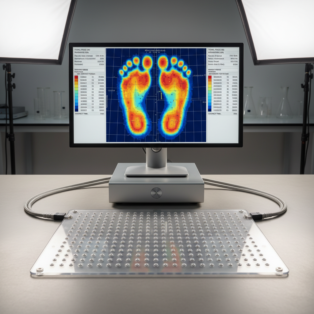
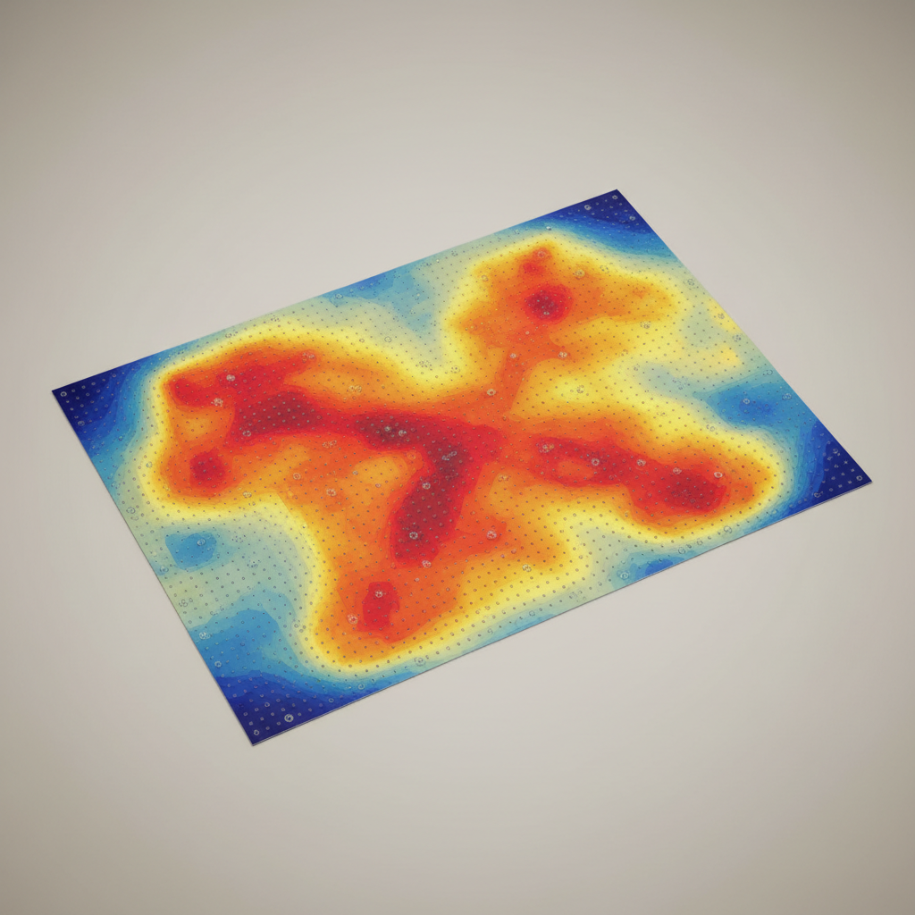
Every scan captures 14 discrete plantar pressure zones — metatarsal heads, arch apex, heel strike, and lateral borders — giving the technician a biomechanical map no visual assessment can replicate.
The average shell delivers 3.2mm of medial arch elevation — precise enough to eliminate the tibial rotation that drives knee pain.
Of referred patients report measurable pain reduction within 21 days of first wear.
From scan upload to finished shell delivered to your clinic — including two rounds of fit adjustment.
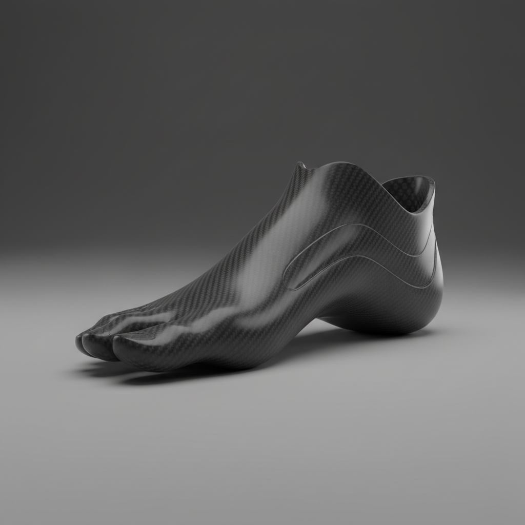
Full carbon fiber reinforcement at 0.4kg. Lighter than EVA alone, with four times the fatigue resistance for high-mileage athletes.
Scan to shell.
Three steps, zero guesswork.
The process a referring podiatrist needs to understand before their first case. Transparent at every stage.
Pressure Scan Upload
Your clinic uploads the pedobarographic scan file directly to our secure practitioner portal. We accept RS-Scan, Tekscan, and Zebris formats.
The scan captures dynamic and static pressure distribution across all 14 mapped zones during a standardized 3-step gait sequence.
Biomechanical Modeling
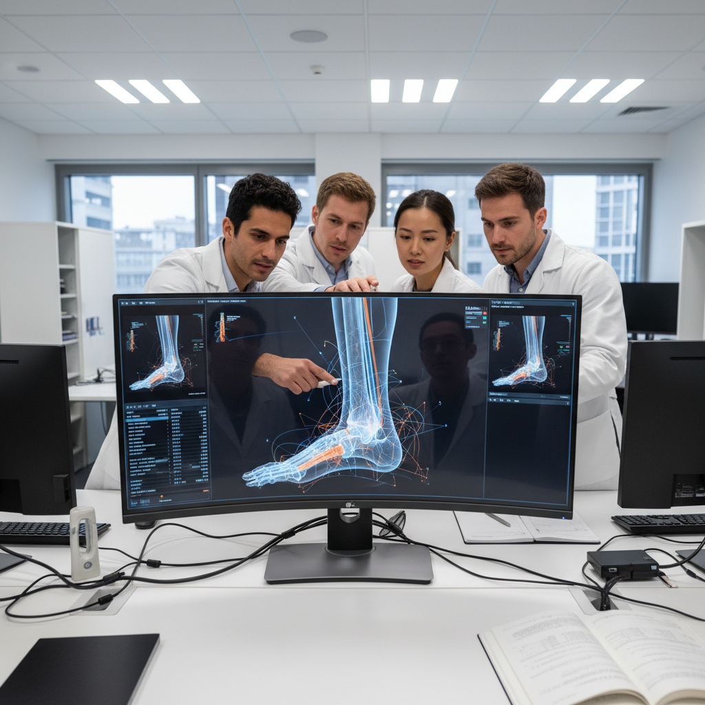
Our technicians translate the pressure map into a three-dimensional correction model — specifying arch height, metatarsal offloading, and heel cup depth to the tenth of a millimeter.
Each model is reviewed by a certified orthotist before carving begins. No algorithm decides the geometry; a trained hand does.
Hand-Finished Shell
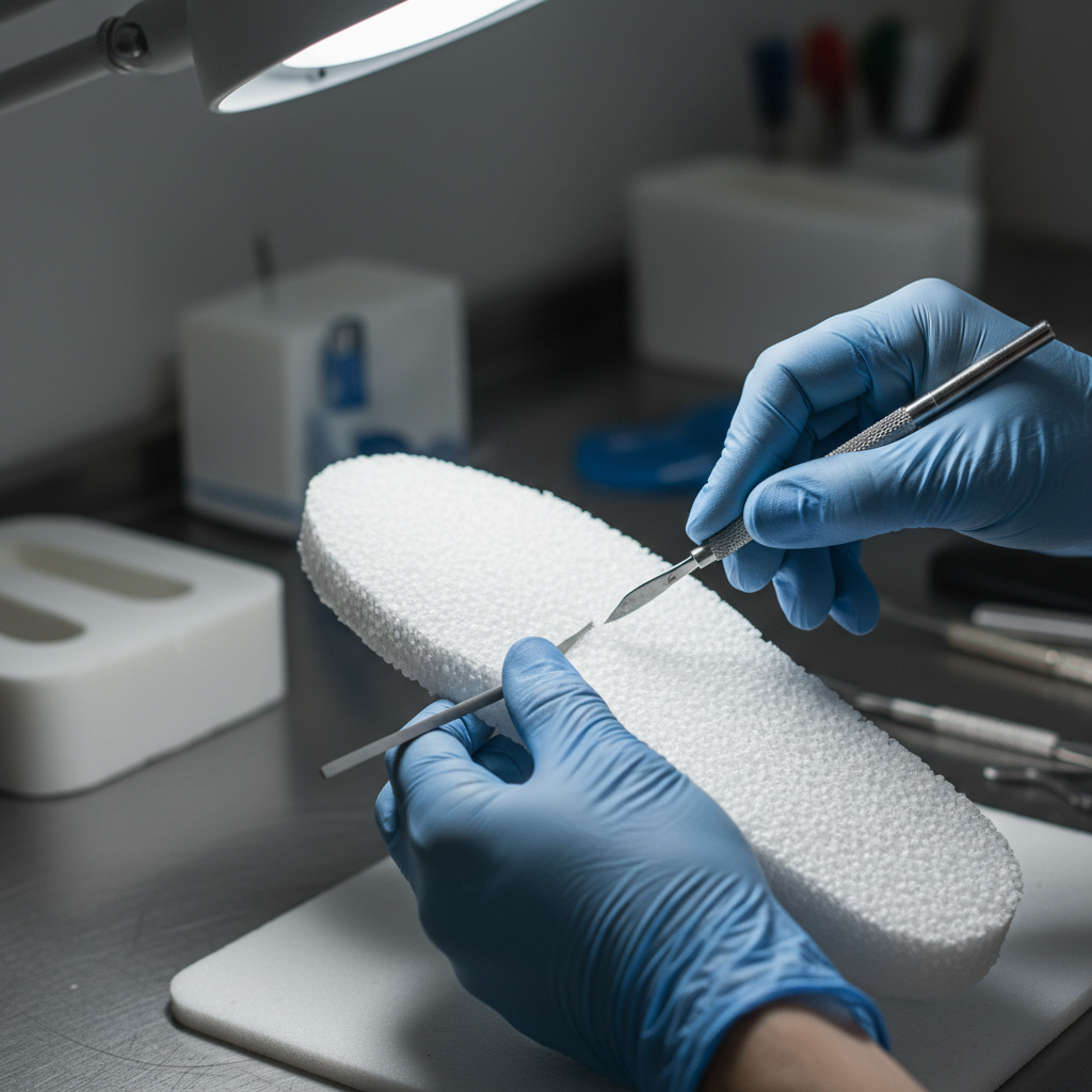
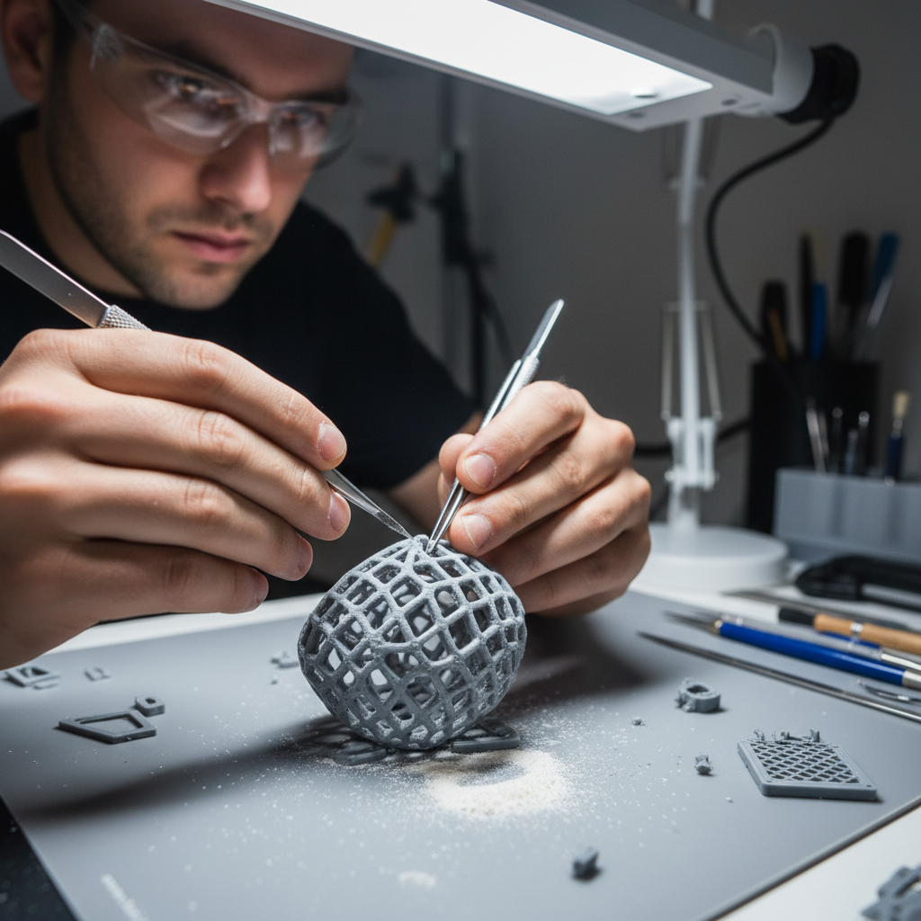
The shell is CNC-roughed from EVA foam or carbon fiber blank, then hand-finished by a technician who has fitted over 300 pairs. The top cover is bonded under pressure.
Carbon fiber shells are post-cured at 120°C for 4 hours. EVA shells are heat-molded to the last. Both are tested under 120kg static load before dispatch.
Three conditions.
One precision answer.
Each guide includes scan parameters, material recommendations, and expected outcomes — ready to reference in a patient consultation.
Plantar Fasciitis
For the marathon runner who has been nursing a tight first step for six months. The orthotic offloads the fascial insertion by redistributing forefoot strike load across the metatarsal heads.
- Fascial tension reduced by avg. 34% on pressure scan
- Heel cup depth calibrated to calcaneal pitch angle
- Compatible with racing flats and stability trainers
Post-Surgical Recovery
After bunionectomy, Achilles repair, or flatfoot reconstruction, the foot has changed. The orthotic is built to the post-operative anatomy — not the pre-surgical one.
Diabetic Ulcer Prevention
The pressure ulcer that precedes amputation forms over weeks of undetected peak loading. The orthotic distributes that load before the tissue breaks down.
The Practitioner
Scanning Guide.
48 pages. The complete clinical reference for scan-to-shell orthotics — written for podiatrists, not technicians. Precise enough to cite in a patient chart.
Download the Guide
For certified podiatrists and orthopedic practitioners. Two fields. No marketing sequence.